Axio Recovery
Cornell DEBUT · Cornell University · 8-Person Team (Project Manager)
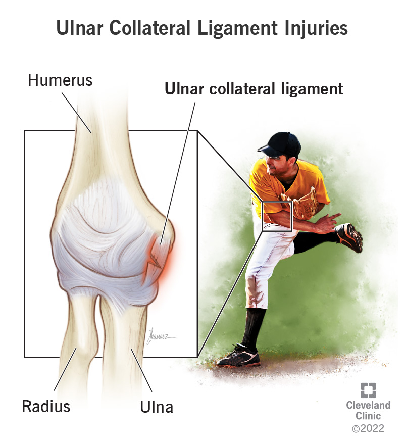
I led an 8-person Cornell DEBUT team from blank schematic to validated diagnostic hardware, a wearable that estimates elbow torque to 2.58% error against motion-capture ground truth.
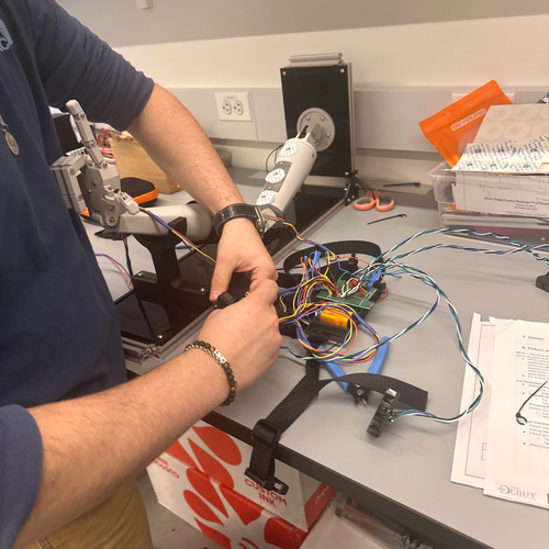
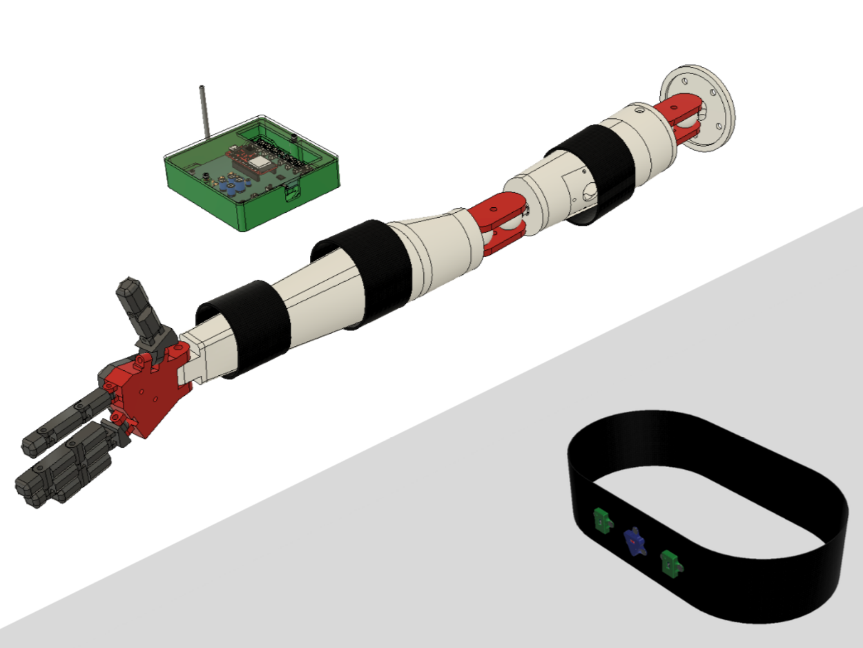
 Final system, V2.0
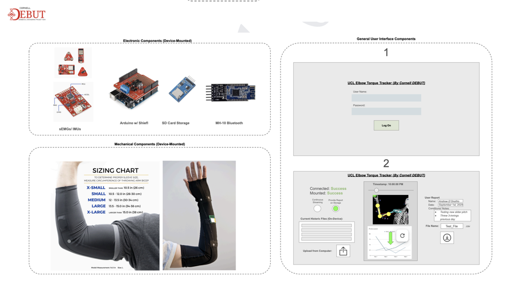
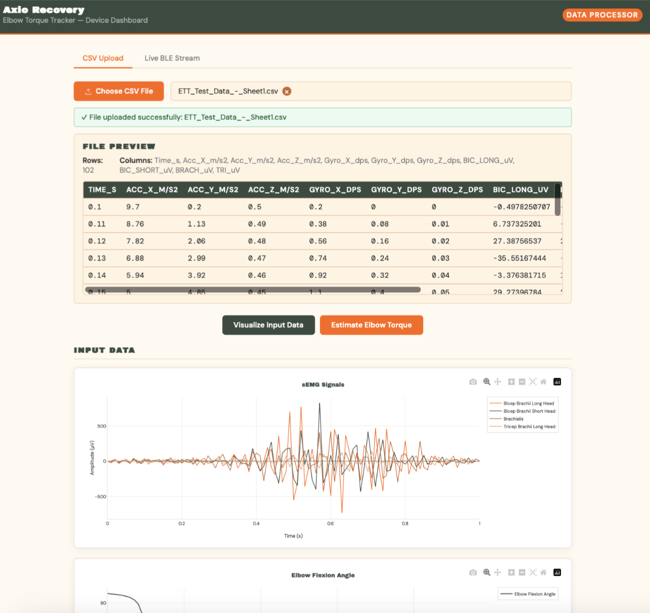 Clinician dashboard: axiorecoverytracker.com
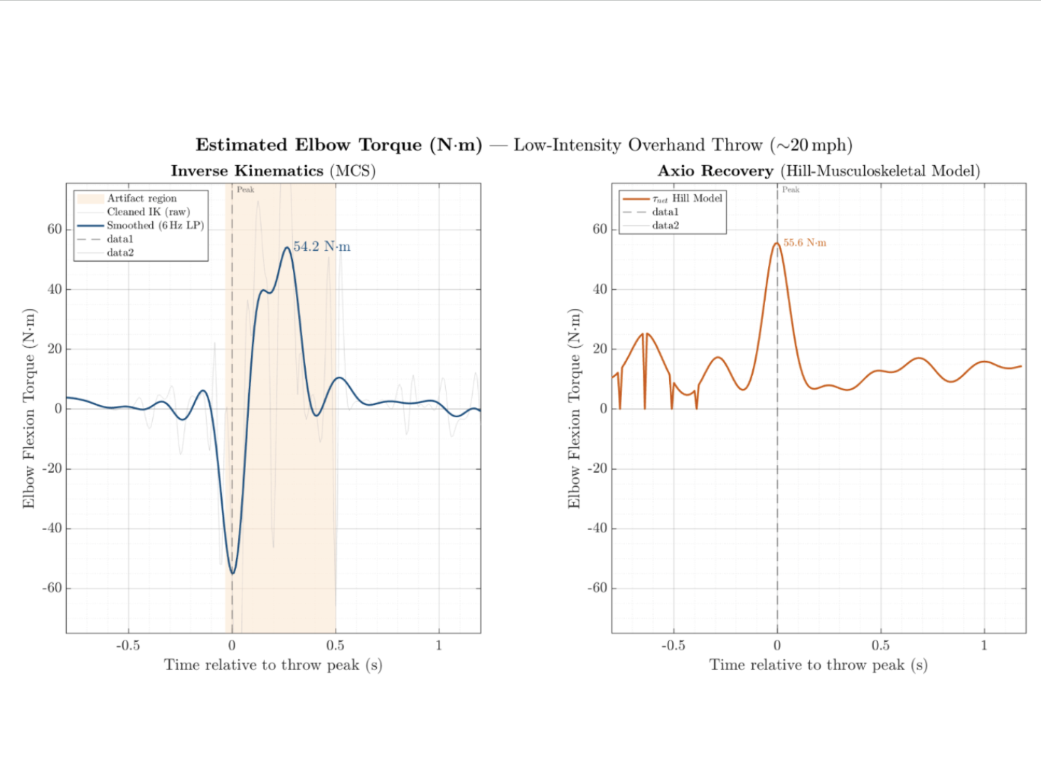 Validation: OptiTrack vs. Axio Recovery
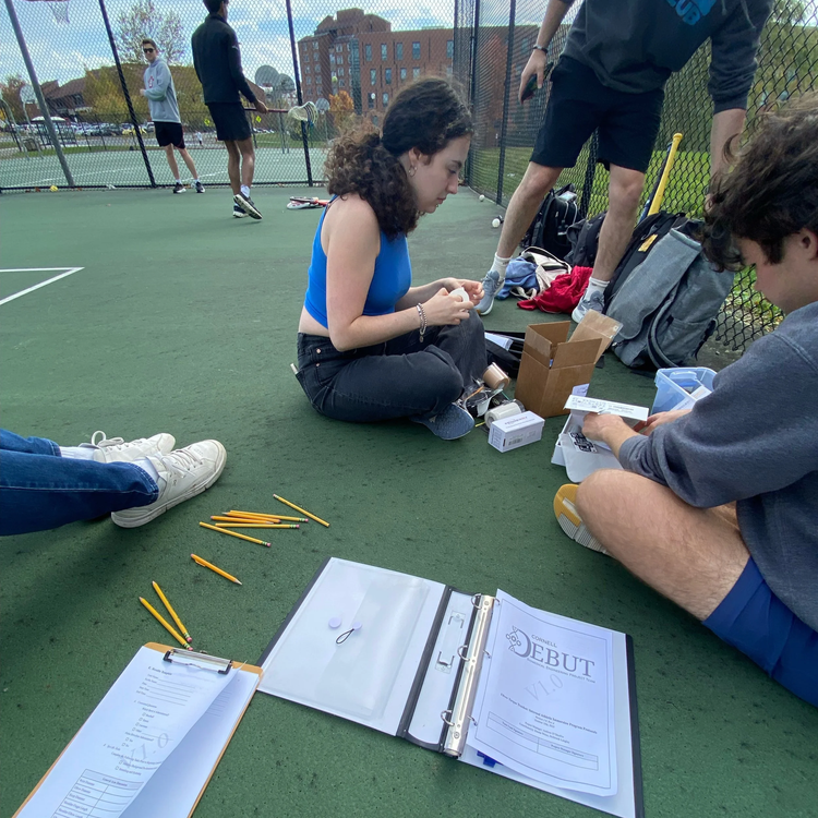
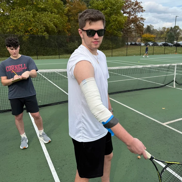
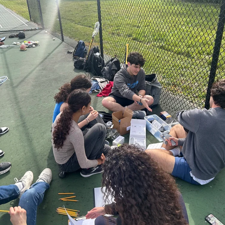 Field testing: throwing motion capture
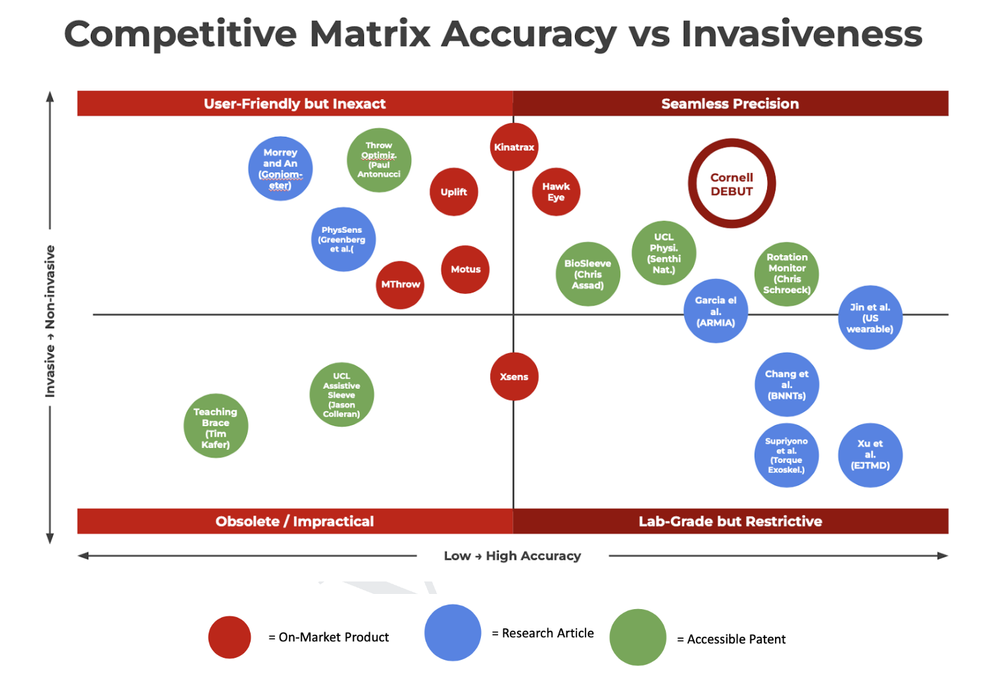 Competitive matrix: accuracy and cost targets vs. Pulse and lab motion capture
Systems Engineering
sEMG / IMU Sensing
Hill's Musculoskeletal Model
PCB Design (KiCAD)
Embedded C++
Python Signal Pipeline
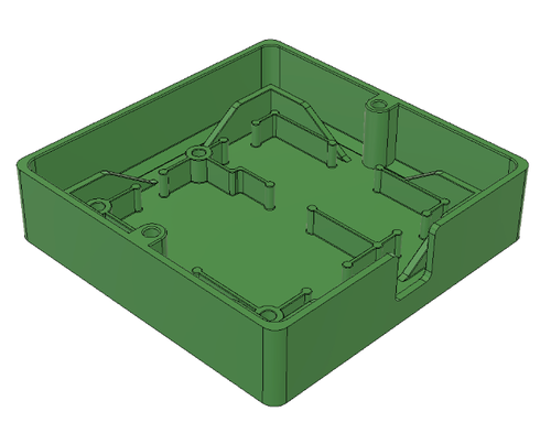
Fusion360
V&V Testing
OptiTrack Motion Capture
GitLab CI
Technical Leadership